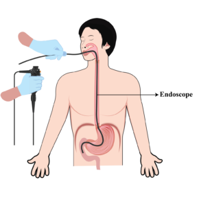
The esophagus is an organ that transfers food from the oral cavity to the stomach and is also known as a food pipe. Esophagus wall is made up of various layers of tissues such as mucosal membrane, muscles, and connective tissue. The inner part of the esophagus is coated by a layer called mucosa. This membrane is exposed to all the foods and harmful substances swallowed through the mouth and passed into the stomach. The mucosal membrane is covered by a special epithelium known as the squamous epithelium. Cancer may develop in the esophagus, which causes narrowing in the lumen of the esophagus and may lead to difficulty in swallowing food.
Types of Esophageal Cancer
There are Two Types of Esophageal Cancer:
- Squamous cell carcinoma: It mostly affects the upper and middle part of the esophagus. It begins in the squamous epithelium that lines the esophagus. This type of cancer is generally associated with factors like tobacco, smoking, consumption of alcohol, and poor dietary habits.
- Adenocarcinoma: This type of cancer is often associated with gastroesophageal reflux disease (GERD) and Barrett’s esophagus where the normal squamous lining of the lower part of the esophagus is replaced by the columnar epithelium cells. The underlying cause of Barrett’s esophagus is chronic exposure of squamous epithelium to acid abnormally refluxed from the stomach in the esophagus. Adenocarcinoma commonly occurs in the lower part of the esophagus. Obesity and smoking has also been associated with adenocarcinoma of the esophagus.


Symptoms of Esophageal Cancer
The early-stage esophageal cancer may not show any signs or symptoms. But as the cancer progresses, the following symptoms become noticeable.
- Difficulty in Swallowing: As the cancer grows in size, the normal passage of food through the esophagus is obstructed, which may lead to difficulty in swallowing. Patients may experience the sensation of food being struck in the esophagus. Initially, difficulty is more for solid foods such as fruit bites and breads, but gradually progresses to semisolid and liquid food also. Dysphagia may occur due to other diseases also, such as Peptic stricture and GERD. Not all patients with dysphagia has underlying cancer.

- Weight loss: due to the difficulty in swallowing the food, the patient may change- eating habits may avoid food also, leading to unintentional weight loss.

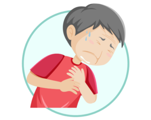
- Pain: Some patients with esophagus cancer may develop chest pain, especially after intake of food.
- Bleeding: Cancer tissue has fragile blood vessels, which may bleed. Patients may have bleed from the mouth or may have black stools.
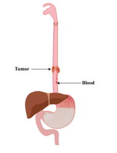
- Coughing or Hoarseness: Cancer tissue may invade the respiratory organs such as larynx, trachea, and lungs, leading to changes in voice and cough.

- Fatigue: As the cancer progresses, it may lead to fatigue and weakness, due to poor dietary intake and release of harmful chemicals by the cancer tissue.

It is important to note that symptoms mentioned here may occur due to non-cancerous conditions of the esophagus also. Patients suffering from the above symptoms may have benign on cancerous diseases of the esophagus also.
Diagnosis of Esophageal Cancer
Early detection and accurate diagnosis are crucial for the best possible outcome in treating esophageal cancer. If after listening to the patient’s complaints and physical examination, esophageal cancer is suspected, investigations are carried out to confirm the disease.
Some of the diagnostics tests are as follows:
- Endoscopy with Biopsy (Esophagogastroduodenoscopy or EGD): Endoscopy is the most important test to diagnose esophageal cancer. Many patients has a fear of passage of discomfort during endoscopy. Endoscopy is performed in fasting condition; the patient should usually avoid food for 8 hours before endoscopy. Most commonly intravenous anaesthesia is given, and endoscopy is performed after proper sedation. Endoscopy performed under anaesthesia becomes a painless procedure without any discomfort. A thin, flexible tube called an endoscope with a camera is passed through the mouth into the esophagus. At the same time, endoscopy can also be passed into organs below the esophagus such as the stomach and duodenum. During endoscopy, a detailed examination of these organs is done, and if cancer is suspected, a biopsy may be obtained. Biopsy for esophagus cancer is a safe procedure without significant complications. Contrary to belief, esophageal cancer does not spread during biopsy sampling.

- Image-enhanced endoscopy: New-generation endoscopes are equipped with new technology such as Image-enhanced endoscopy. This technique can diagnose esophageal cancer at very early disease. Cancers diagnosed in such an early stage may be curable with endoscopic resection techniques such as Endoscopic mucosal resection and endoscopic submucosal dissection.
- CT Scans: In patients who are not fit for endoscopy, a CT scan may be performed. However, CT scans may miss early-stage cancers.

- Barium swallow: Barium swallow is a preliminary test in patients with symptoms of dysphagia and may miss early-stage cancers. During this test, the patient swallows a special liquid containing Barium, and X ray is obtained. Based on barium swallow, cancer or non-cancerous diseases are suspected, and patients are investigated accordingly.
Staging of cancer: Once the cancer is suspected, various tests are performed to stage the disease.
Principle of cancer staging: Staging is based on tumour spread and based on three parameters.
- Depth of invasion in the esophagus and surrounding organs such as traches, larynx, heart, and lungs. Staging based on tumour depth is known as T staging.
- Involvement of lymph nodes. Lymph nodes are small glands around body tissue. The function of lymph nodes is to carry the remains of blood known as lymph from tissue to the heart. Lymph nodes without involvement by cancer tissue are not enlarged and are not seen on CT scans. Staging based on lymph node involvement is known as N staging.
- Cancer may spread to distant organs such as lungs and liver through blood. Staging based on spread to other organs is known as M staging.
Staging of Esophageal Cancer
CT Scan or PET CT Scan: During a CT scan, the depth of invasion of esophageal cancer along with its spread to lymph nodes or other organs can be assessed. Advanced stages of cancer can be diagnosed with a CT scan. If CT shows involvement of lymph nodes or peri esophageal tissue, chemotherapy and radiotherapy will be required before surgery. If CT is showing spread to other organs such as lungs and liver, surgery is avoided in these patients and
palliative chemotherapy is advised.
Endoscopic Ultrasound (EUS): Endoscopic ultrasound is an endoscopy-based test. A special endoscope is used for this test, where an ultrasound probe is attached to the tip of the endoscope. This test has a special role if a CT scan is suggestive of less danced disease with non-involvement of periesophageal tissue, lymph nodes, or distant organs, EUS is performed in these patients. EUS provides excellent information about the layers of the esophagus and lymph nodes around the esophagus. If EUS shows involvement of tissue around the esophagus and lymph nodes, these patients will require chemotherapy or radiotherapy prior to surgery.
Image-enhanced Endoscopy: Image-enhanced endoscopy is a special technique used to confirm very early cancers that can be removed with endoscopic resection techniques such as endoscopic mucosal resection (EMR), and endoscopic submucosal dissection(ESD). New-generation endoscopes are equipped with narrow-band imaging, and tests can be performed while performing the first endoscopy. If the tumor is confined to the upper 2/3 of the mucosal layer, such a tumor can be removed with endoscopic techniques.
Based on all these investigations, cancer is staged and treated accordingly.
Treatment Options
Treatment of cancer depends upon the stage and site (upper or lower esophagus)
Following are some general treatment plans for esophageal cancer:
Very early esophageal cancer: Tumor confined to the upper 2/3 of the mucosal layer without involvement of lymph nodes or distant organs are treated by endoscopic resection techniques such as
Endoscopic Submucosal Dissection (ESD): It is a minimally invasive endoscopic procedure that can be used to manage very early stages of cancer. In this procedure, an endoscope is inserted through the mouth and the doctor’s tumor is dissected from the underlying layers of the esophageal wall. Large tumor can also be removed with minimal damage to the surrounding healthy tissue.
Endoscopic Mucosal Resection (EMR): This procedure is used to remove superficial tumors, which are small in size. A snare is passed through the endoscope to remove the tumor. Other treatment options include Surgery (surgical removal of the tumor), Radiotherapy, Chemotherapy and Immunotherapy.
Early-stage cancer: If tumor has gone beyond the deep mucosa, but is confined to esophageal wall, without involvement of. Surrounding tissue, lymph nodes, and distant organs, such cancers can be considered as early stage cancers. Further treatment depends on the location of the cancer. Tumor in the upper esophagus are treated by chemotherapy and radiotherapy and cancers in the lower esophagus can be treated by surgery.
Advanced cancer: Tumors involving periesophageal tissue and lymph nodes are considered as advanced cancers. Tumor in the lower esophagus are treated by chemotherapy and radiotherapy, followed by surgery. Tumor in the upper esophagus are treated by chemo-radiotherapy without surgery.
Very advanced disease: Tumor involving other organs such as the liver and lung are treated by palliative chemotherapy depending upon the condition of the patient.
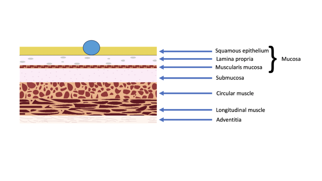
Tumor confined to epithelium, T1a, m1, if no lymph node or distant metastasis, endoscopic resection

Tumor confined to lamina propria, T1a, m2, if no lymph node or distant metastasis, endoscopic resection
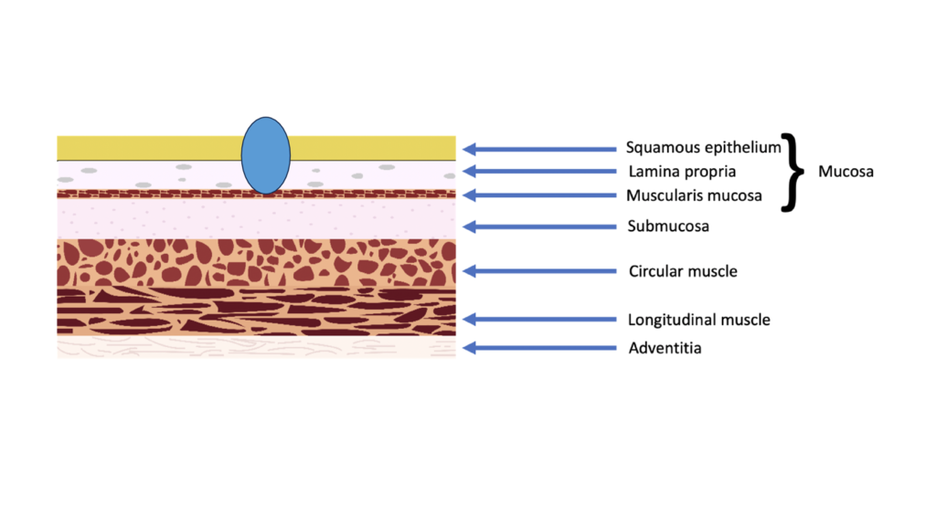
Tumor confined to muscularis mucosa, T1a, m3, if no lymph node or distant metastasis, endoscopic resection or surgery
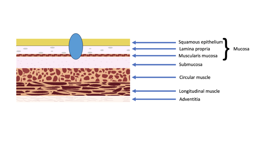
Tumor confined to superficial 1/3 of submucosa, Tba, SM1,
If no lymph node or distant metastasis, endoscopic resection or surgery
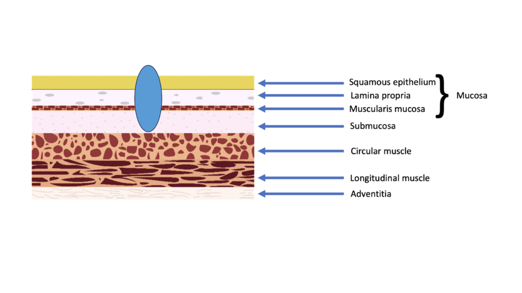
Tumor invading deep submucosa T1b, SM2 or SM3
If no lymph node or distant metastasis, surgery in distal esophagus
Chemotherapy and radiotherapy in proximal esophagus

Tumor invading muscularis propria T2
If no lymph node or distant metastasis, surgery in distal esophagus
Chemotherapy and radiotherapy in proximal esophagus
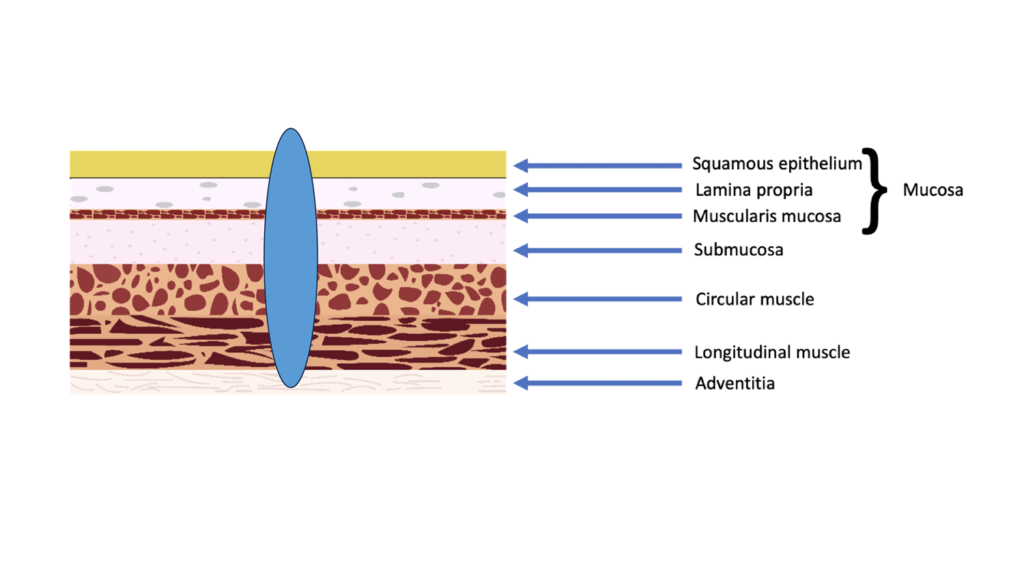
Tumor invading adventitia T3
Chemotherapy and radiotherapy followed by surgery in the distal esophagus
Chemotherapy and radiotherapy in the proximal esophagus
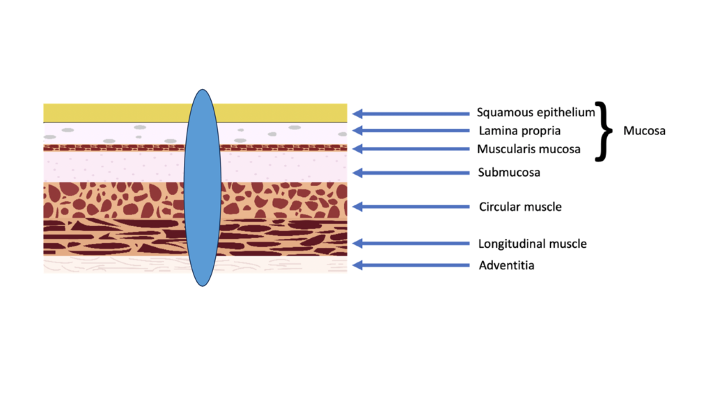
Tumor invading beyond adventitia T4
Chemotherapy and radiotherapy followed by surgery (if feasible) in distal esophagus
Chemotherapy and radiotherapy in the proximal esophagus
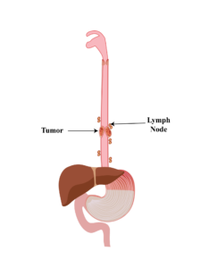
Tumor in lymph Node
Chemotherapy and radiotherapy in proximal esophagus
Chemotherapy and radiotherapy followed by surgery in distal esophagus
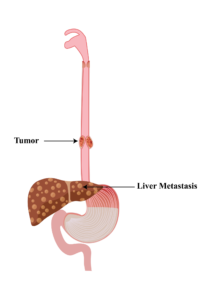
Role of Esophageal Stenting in Carcinoma Esophagus
For patients who have advanced diseases, a cure of disease may be difficult. In these patients, the metal stent may be placed in the esophagus across the growth for palliation of symptoms of dysphagia.
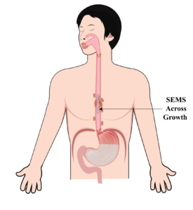
Prevention of Esophageal Cancer
We can reduce the risk of development of esophageal cancer by adopting a healthy lifestyle and minimizing exposure to certain potential risk factors.
- Maintaining a healthy weight: Maintaining a healthy weight by eating a balanced diet and doing regular exercise can help to reduce the risk of developing cancer as obesity is a risk factor for esophageal cancer.

- Proper Diet: A healthy diet can help reduce the risk of esophageal cancer and other diseases. A diet must be rich in fruits and vegetables, which have antioxidants and micronutrients, which may help prevent cancer.

- Avoid tobacco, smoke, and limit alcohol: Consumption of these factors is associated with an increased risk of esophageal cancer. Stopping tobacco and smoking, and limiting the use of alcohol may prevent esophageal cancer.

- Screening for esophageal cancer: Regular screening for individuals with high-risk conditions such as Barrett’s esophagus may reduce the risk of esophageal cancer.

